flow volume loop
One of the more recognizable flow-volume loop contours is the one associated with severe airway obstruction. Characteristic flow-volume loop patterns are.

Pin On Respiratory Therapy
A graph of the rate of airflow as a function of lung volume during a complete respiratory cycle consisting of a forced inspiration followed by a forced expiration.
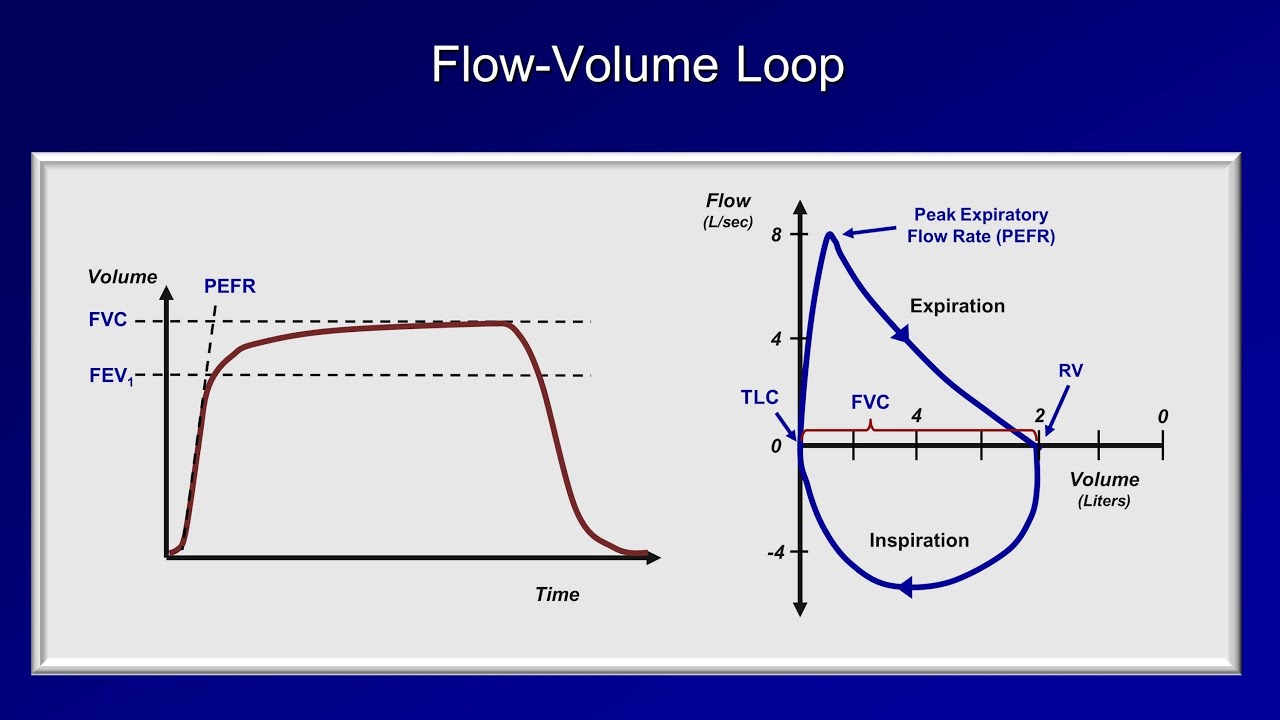
. Normal is 070 or greater. A flow-volume loop which graphically depicts the rate of airflow on the Y-axis and the total volume inspired or expired on the X-axis. At the start of the test both flow and volume are equal to zero. After the PEF the curve descends the flow decreases as more air is.
Indeed both the LITFL entry on flow-volume loops and the AnaesthesiaUK revision article use the formal pulmonary function test standard of flow-volume loop interpretation. Flow volume loop. Flow Volume Loops. Obtaining a flow volume loop in the pulmonary function laboratory is a simple and effective method of noninvasively evaluating a patient for the presence of an upper airway obstruction.
Inspiratory limb of loop is symmetric and convex. The plotted curve appears as a loop and is used in assessing pulmonary function. In obstructive lung disease such as COPD the expiratory limb takes on a coved shape Figure 1b. Flow volume loopBronchodilator response Dr.
A flow-volume loop plots flow on the y axis and volume on the x axis. The values of the parameters are higher than 80 of the predicted values while the Tiffeneau index FEV1FVC100 is higher than 70. So in 1 second how much are you able to expel. Expiratory limb is linear.
For example airflow is normally slower at low lung volumes because elastic recoil is lower at lower lung volumes. Flow rates at the midpoint of the inspiratory and expiratory capacity are often measured. Analyzing flow volume loops provides a quick way to detect lung disease. It is also important to keep in mind that this graph truly is a loop that is cyclic in nature ie.
Changes in the contour of the loop can aid in the diagnosis and localization of airway obstruction 1. Common problems that can be diagnosed by analyzing Ventilator waveforms Abnormal ventilatory Parameters lung mechanics Eg. The inspiratory part of the loop is shaped like a half circle. Give 2 examples of and be able to identify the flow volume loop characteristics of 2 factitious abnormalities.
The flow-volume loop is a test of ventilatory function in which a graphic recording of maximal flow rates is displayed at all lung volumes during a maximal effort forced expiratory and inspiratory vital capacity maneuver. Spirometry tests were originally recorded by a pen trace on kymograph paper as a volume-time VT curve. After the starting point the curve rapidly mounts to a peak. Provide a graphical analysis of inspiratory and expiratory flow from various inspired lung volumes.
TLCLung partition Hyperinflation Dr. The graph represents a classic example of a flow volume loop Looking at the example above now we can start talking specifics. Pulmonary diseases produce characteristic flow-volume loop abnormalities. Patterns of pulmonary function tests PFTs and flow-volume loops among patients with clinically important tracheobronchomalacia TBM are not well described.
The basic forced volume vital capacity FVC test varies slightly depending on the equipment used either closed circuit or open circuit but should follow the ATSERS Standardisation of Spirometry. Small studies suggest 4 main flow-volume loop morphologies. Flow volume loops. Patients with pulmonary fibrosis have low lung volumes and their airflow appears to be decreased if.
Maximal inspiratory flow at 50 of forced vital capacity MIF 50FVC is greater than maximal expiratory flow at 50 FVC MEF 50FVC because. A normal flow volume loop Figure 1a has a characteristic shape. Physiology lecture on respiratory physiology - Hypoxia details the causes of various types of hypoxia ie hypoxic hypoxia anemic hypoxia ischemic hypoxia a. Loops are representations of pressure vs volume or flow vs volume.
The flow-volume loop is a plot of inspiratory and expiratory flow on the Y-axis against volume on the X-axis during the performance of maximally forced inspiratory and expiratory maneuvers. The appearance of the loop can reveal the presence of obstructive andor restrictive lung disease. Along with spirometry a flow-volume loop is also typically generated. Most of what is written about flow-volume loops refers to these.
We studied common PFT and flow-volume loop. The location of the airway obstruction can also be. What is the volume of air expelled in 1 second. Fixed lesions cause plateaus in both the inspiratory and expiratory limbs of the flow volume loop.
Overdistension Auto PEEP COPD Patient-ventilator Interactions Eg. Specifically this type of loop shows an abrupt decrease in flow rate following the peak flow with a more gradual decrease in flow rates during the remainder of the exhalation. A normal Flow-Volume loop begins on the X-axis Volume axis. Flow-volume FV loops extFVLs obtained during exercise and plotting them according to a measured end-expiratory lung volume EELV within the maximal FV envelope MFVL more specific information is provided on the sources and degree of ventilatory constraint.
Flow-volume loop FVL contours however are. A normal flow-volume loop. Chandrasekhar PFT FVCBronchodilator response Dr. The test is easy to demonstrate administer and analyze.
Because the changes in the contour of a volume-time curve that occur with various lung disorders are very subtle it is a less than ideal tool for interpreting results. A normal expiratory flow-volume loop has a triangular shape with its top at the left. Breathing across a pneumotachograph subjects inhale to TLC - FEC manoeuvre - rapidly inhale back to TLC. The principal advantage of the flow-volume loop is that it can show whether airflow is appropriate for a particular lung volume.
Low maximum forced expiratory flow biphasic expiratory curve flow oscillations and notching. Chandrasekhar PFT TLCPartition Normal Dr. N Loops other possibilities 38 n Flow-volume loop 38 Trends reviewed 40 n Documentation of a weaning process 41 n Lung parameters based on peak and plateau pressure 43 Capnography keeping an eye on the details 44 n The physiological capnogram 46 n Interpretations of capnograms 47. Things begin at the origin of the graph travel in the positive XY axis directions as the patient is exhaling then reverse in a route back to the origin to represent.
Typically when one thinks of flow-volume loops one refers to the classical loops of forced expiratory spirometry. Chandrasekhar PFT Flow volume loop Normal Dr. Flow volume loop Normal Dr.

Medicowesome Flow Volume Loop Notes And Mnemonics Respiratory Therapist Student Mnemonics Respiratory Therapy

Pin On Education Lung

Pin On Usmle Step 1

Schematic Flow Volume Loop Configurations In A Spectrum Of Airway Lesions A Is Normal B Is Variable Extrathoracic Upper Ai Upper Airway Immunology Pediatrics

Pin On Respiratory Therapy

Pulmonary Function Icu Nursing Medical Jobs Respiratory Therapy
Posting Komentar untuk "flow volume loop"